Neuropathological Autopsy Technique
The neuropathological autopsy comprises two large chapters. The first is the cranial autopsy and the second is the spinal autopsy. In the first, the brain is released and in the second, the spinal cord and then proceed to its study.

Neuropathological autopsy is a procedure that is carried out to identify possible lesions in the nervous system that lead to death. These injuries can be of a primary or secondary nature.
Primary injuries are those that correspond to the fundamental or direct process that leads to death, while secondary injuries are those that participate in another fundamental pathology present in the body.
In the neuropathological autopsy, extraction and sampling of the nervous system are carried out. These processes are regulated and vary in quantity and location, depending on whether the lesions are of a primary or secondary nature.
The macroscopic neuropathological autopsy technique comprises two major phases: the cranial autopsy and the spinal autopsy. In the cranial autopsy, the extraction and study of the brain is carried out. In the spinal autopsy, the same is done with the spinal cord, roots and posterior nodes.
Steps in neuropathological autopsy
Brain removal

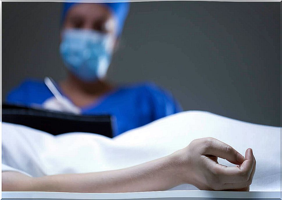
It is recommended that the brain be removed within 24 hours of death. Generally, those who perform this procedure are a pathologist, a technician, and an assistant. The steps to remove the brain are as follows:
- Location of the corpse on the autopsy table in the supine position. The neck and occiput should be located in a headrest, in order to elevate the skull and facilitate maneuvers.
- A scalpel incision is made from one pinna to the other. The depth must reach the periosteum, that is, very close to the bone.
- Subsequently, the periosteal-cutaneous planes are separated, taking them backwards and forwards. With this, the skull is exposed.
- A circular saw cut is made, starting at the front of the skull and going around until reaching the same starting point. The cut should not extend beyond the dura.
- Superior longitudinal sinus opening. It opens from front to back. A small part of the dura is taken and then cut laterally. The brain must be exposed.
- The area of the front poles continues to be separated. The best technique is to do it with the index and middle fingers using gentle tugs. Then the optic chiasm and the remaining parts of the cranial nerves are cut. The brain must be free.
- The two sides of the tentorium or cerebellum are cut with the scalpel.
- The brain, cerebellum, and trunk are gently pulled. Then, the bulb is cut with a scalpel through the foramen magnum. The cut-off must be very low to obtain the complete sample of the medulla oblongata. The brain must be completely released.
- The posterior clinoid processes are ruptured with the chisel. The sella turcica is then enlarged with the hands and the pituitary gland is removed with forceps and a scalpel.
Management of the brain in neuropathological autopsy

After the brain is removed, it must be weighed on a scale. Then it must be suspended with a thread that goes from the basilar to the brainstem. Next, the brain is floated in 10% formalin.
The container must be hermetically closed and labeled. It is normally left there for 15 days, except in cases where prion disease is suspected. If that’s the case, it should be left for a month, at least.
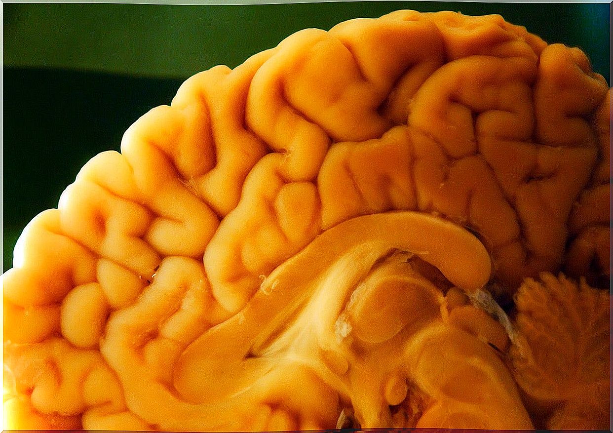
Once this period has elapsed, the external macroscopic study is carried out. The first step is to wash the brain in water for 24 hours. Then palpation and inspection should be carried out. Then the coronal cuts are made and the samples are taken for later microscopic study.
Spinal autopsy
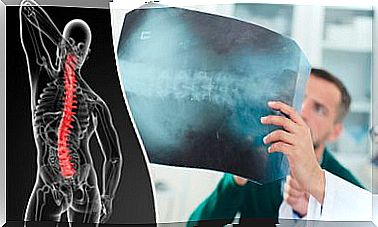
The other great chapter of the neuropathological autopsy is the spinal autopsy. In this, the first thing that is done is to extract the spinal cord, through an internal or external approach. Once released, it is ready for palpation and inspection on both sides.
Next, the spinal cord is sampled. To do this, cross-sectional cuts are made, every 3 or 4 centimeters. If there is a specific spinal pathology, representative samples of the lesion should be taken. Samples should be taken from the cervical, dorsal, and lumbar regions. Also on the sacral level, when possible.


